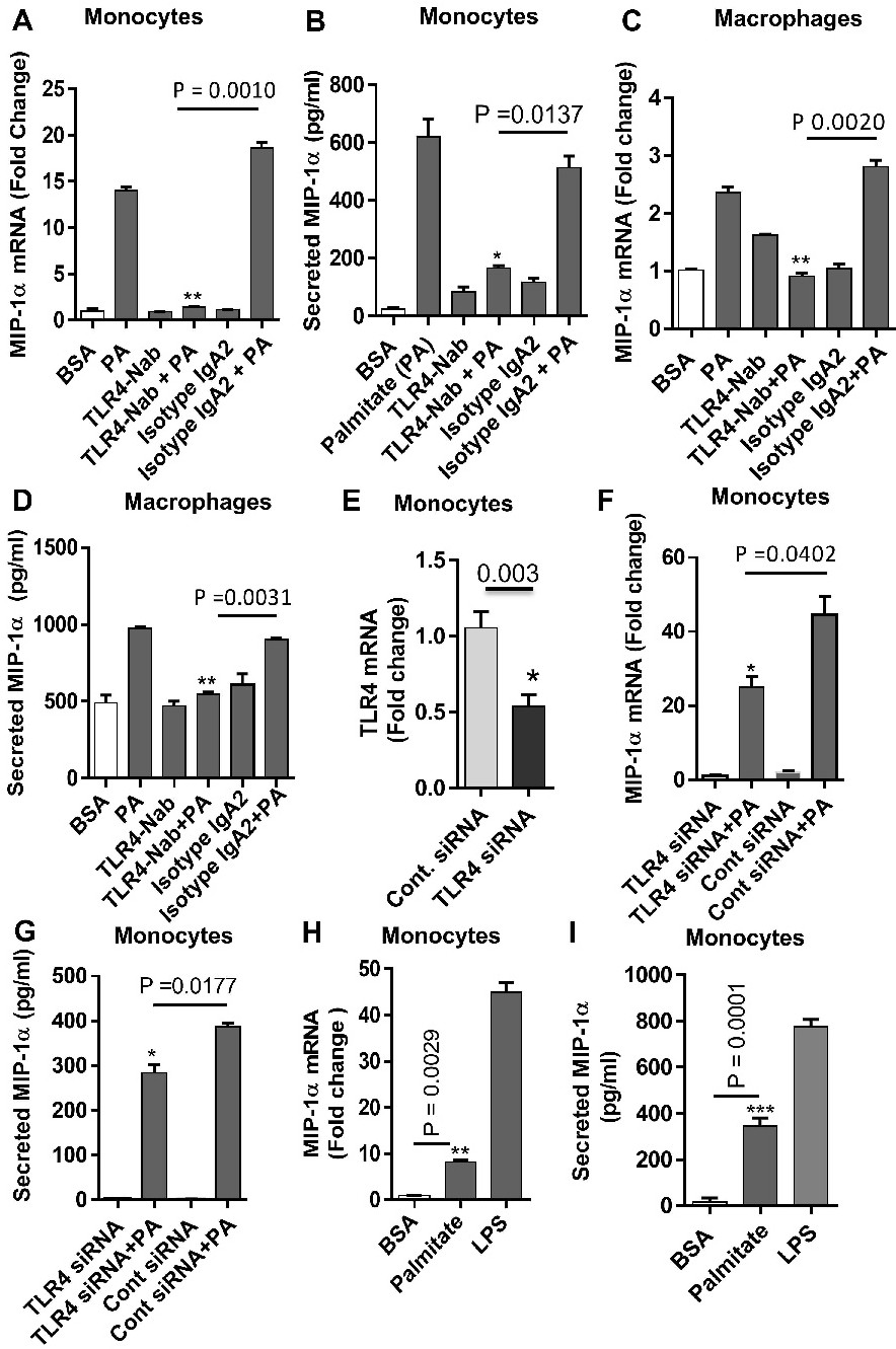
Fig. 3. Inhibition of TLR4 down-regulates the palmitate induced MIP-1α. Anti-TLR4 antibody or isotype-matched control antibody (IgA) treated THP-1 cells were incubated with 0.1% BSA (vehicle) or palmitate (100μM) for 24 hrs. Cells and culture media were collected. MIP-1α mRNA and protein were determined (A and B). THP-1 derived macrophages were treated with TLR4 and control antibody and treated as indicated. MIP-1α mRNA and protein were determined (C and D). THP-1 cells were transfected with either control siRNA (30nM) or siRNA (30nM) against TLR4 (E). Cell were treated as indicated and MIP-1α expression was determined (F and G). THP-1 cells were incubated with 0.1% BSA (vehicle) palmitate (100μM), LPS (10 ng/ml; a positive control for TLR4 activation) for 24 hrs. Cells and culture media were collected. MIP-1α expression was determined (H and I). The data are presented as mean ±SEM. Statistical analysis was done using t-test. P<0.05 was considered as statistically significant (* P<0.05; **P< 0.005).